
A visceral-fat estimate from DXA is not automatically the same quantity as one from MRI. For Bali readers researching tesamorelin, the useful first step is to identify the method and body region on the report. Similar labels can conceal different measurements, even when both refer to abdominal fat.
What a recent comparison found
A 2026 DXA–MRI study examined 50 matched scan pairs from 28 adults with obesity and without diabetes. The researchers matched an L3 region. The methods were strongly correlated, but DXA estimates were systematically higher in that dataset and absolute agreement was limited.
That is not a universal correction rule. It describes those participants, methods and analyses. Fifty scan pairs also do not mean fifty different participants; retaining the participant count avoids making the study appear larger than it was.
Check these report fields
| Field | What to retain | Why it matters |
|---|---|---|
| Method | DXA, MRI or CT | The technologies differ |
| Region | Exact anatomical definition | Different regions are different quantities |
| Units | Area, volume or another output | Numbers cannot be compared by label alone |
| Analysis | Software and report details | Processing influences the estimate |
If a report was supplied as a cropped screenshot, ask for the complete document before comparing it with an older scan. A percentage or score without its method and region can be difficult to interpret meaningfully.
Keep tesamorelin research separate
The EGRIFTA WR label describes product-specific research in HIV-associated lipodystrophy. Its pivotal trial endpoint used CT visceral-fat assessment at L4–L5 after 26 weeks. That is not the same protocol as a general DXA body-composition report or the L3 MRI comparison above.
The tesamorelin listing and shop identify the catalogue format. They do not establish equivalence to EGRIFTA WR or determine whether imaging is appropriate for an individual. This article explains reports rather than recommending a scan or a treatment.
FAQ: Report questions
Can I compare two “visceral fat” numbers directly?
Only after checking what each actually measures; matching labels alone is insufficient.
Does correlation mean equal values?
No. Methods can move together while giving systematically different estimates.
Did the tesamorelin trials use this DXA comparison?
No. The label's CT L4–L5 endpoint is a different research measurement.
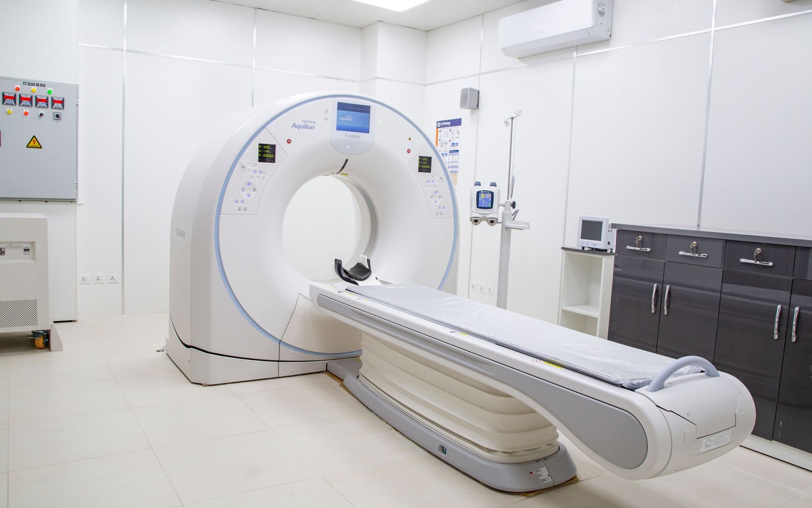
Photograph: contact me +923323219715 · Pexels. Medical imaging equipment, illustrating the distinction between measurement methods. Illustrative stock photograph; it does not show the study preparation or its results.
Sources and further reading
- Comparison of MRI and DXA for visceral adipose tissue quantification in obese adults without diabetes — Mancilha et al., Journal of Clinical Densitometry. Source checked 2026-10-27.
- EGRIFTA WR prescribing information — DailyMed. Source checked 2026-10-27.